Showing posts with label VDJ recombination. Show all posts

antibodies

Antibodies are glycoproteins of the immunoglobulin superfamily, and are adhesion-signaling molecules that recognize (bind to) specific antigens. Antibodies are synthesized by B cell-derived plasma cells.

▼: adhesion molecules : antigen binding site : C : CH1-4 : cellular adhesion molecules : complement fixation : complementarity determining regions : constant domains : domains : evolution of immunoglobulins : Fab : Fc : heavy chain : hinge region : Ig supergene family : isotypes : kinase activation : light chain : location of Ig classes : membrane-bound Igs : multimeric structures : tissue location : V : VDJ recombination : VH, VL : variable domains :▼
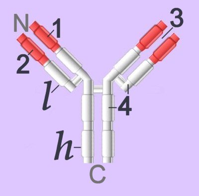
Immunoglobulins (left - click to enlarge) comprise two heavy (h) and two light-chain (l) protein subunits, each of which folds into domains (4 on heavy, 2 on light). These adhesion sites or domains contain one or more folds of 60 to 100 amino acids.

Depending upon the character of the heavy chain, immunoglobulins are divided into five classes – IgG, IgD, IgE, IgA, IgM – that are expressed in different tissues. The classes are further subdivided into isotypes, which have different properties in terms of complement fixation and binding to immunoglobulin (Ig) receptors.

Members of the immunoglobulin supergene family are found as:
● membrane-bound surface receptors of immune-system cells,
● cellular adhesion molecules, or
● soluble antibodies (γ-globulins) synthesized by activated B cells.

Membrane-bound Igs have a transmembrane segment and a cytoplasmic C-terminal tail. The 2 β- chains are stabilized into sandwiched β sheets that are adherent by virtue of hydrophobic interactions between disulphide bonds. Igs assume a Y-shaped structure "topped" at the extracellular N-terminals by variable domains (red), with a variable domain at the tip of the heavy chain (1) and the light chain (2), between which lies an antigen binding site (3). The variable regions are coded by pluripotential DNA sequences that can generate thousands of polypeptide sequences capable of adhering to millions of different ligands. Binding is homophilic or heterophilic, including binding to different Igs and to integrins. Both light and heavy chains contain constant domains (white, 4).
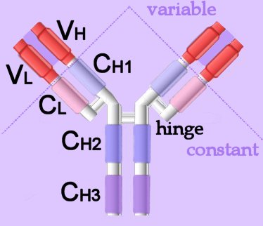
Right - click to enlarge - the heavy chains of IgA, IgD and IgG each have four domains, where those at the N-terminal are variable (VH) and the other three are constant (CH1-3). IgE and IgM have one variable and four constant domains (CH1-4) on the heavy chain. The variable domains are termed Fab, while the constant domains are termed Fc.

The light chains have two domains, one variable domain (VL) at the N-terminal, and one constant (CL) domain.

The antigen binding site lies between VH and VL (shaded lavendar). Most variability is found in three superficial-loop forming regions in the VH and VL domains, which are the complementarity determining regions or CDRs. CDR3 binds antigens and CDR1-2 bind MHCs. CDR3 shows more variation that do either CDR1 or 2.

The domains have related amino acid sequences that possess a common secondary and tertiary structure. This conserved structure is found frequently in proteins involved in cell-cell interactions and is particularly important in immunology. The constant (Fc) regions have complement fixing and Ig receptor binding activity. The hinge region, in IgG, IgA and IgD, is an important sequence of 10-60 amino acids between CH1 and CH2 that confers flexibility on the molecule.

animations Џ B cell selection Џ ELISA test +ve, -ve Џ IgG rotating x- y- axes Џ Rotating mouse IgG2a Molecule (y-axis) Џ somatic recombination of Ig gene Џ spinning IgG1 Kol Џ unfolding (small) IgG . unfolding (large) IgG .

Immunoglobulins attain their enormous variability by splicing components (VDJ recombination) coded in widely scattered sequences of DNA that are located in two different chromosomes. Antigen binding takes place at the heavy chain, which displays enormous variation by virtue of combining 1 of 400 possible variable gene segments with 1 out of 15 diversity segments and 1 out of 4 joining segments. This alternative splicing generates 24,000 possible combinations for the DNA encoding the heavy chain alone. The variable coding segments are assembled together with those for the constant-C segments of the heavy-chain molecule.

Tissue location:
IgA – mucus – gut, respiratory tract
IgD – antigen receptor on B cells
IgE – mast cells – releases histamines in response to allergens
IgG – primary immunity against invading pathogens
IgM – early B cell-mediated response to invading pathogens

Some antibody classes form multimeric structures – pentamers (IgM) and dimers or trimers (IgA). These two isotypes also associate with a small protein called the joining (J) chain required for stabilisation of the complexes.

The immunoglobulin superfamily is evolutionarily ancient, is widely expressed, and is constitutive or long-term up-regulated. Immunoglobulin antibodies are released by activated B cells of the immune system, on which they also act as surface marker proteins. Adherence of immunoglobilins to foreign substances or to cellular invaders may be sufficient to disarm the invader, or the attached antibodies function as attack signal to macrophages and natural killer cells. Adhesion molecules of the immunoglobulin supergene family, activate specific kinases through phosphorylation, resulting in activation of transcription factors, increased cytokine production, increased cell membrane protein expression, production of reactive oxygen species, and cell proliferation.

▲: adhesion molecules ~ adhesion molecules ф antigen : antigen binding site ф APCs ф B cells : C : CH1-4 : cellular adhesion molecules : complement fixation ф complement system : complementarity determining regions : constant domains : domains : evolution of immunoglobulins : Fab : Fc : heavy chain : hinge region ф humoral immunity : Ig supergene family ~ immunoglobulins : isotypes : kinase activation ♦ kinases : light chain : location of Ig classes : membrane-bound Igs : multimeric structures ф receptors ф signaling ф surface receptors ф T cells : tissue location ~ tyrosine kinases : V : VDJ recombination ф VDJ recombination : VH, VL : variable domains :▲

Tables  Fc receptors  Immune Cytokines  Immunoglobulins  Cell Adhesion Molecules  Cell signaling  Receptor Tyrosine Kinases (RTKs)  Receptor Signal Transduction  Second Messengers 

▲ Top ▲

tags

B cells

B cells are lymphocytes (WBCs) that participate in humoral immunity by producing antibodies in response to antigen stimulation.

▼ activation : B-1 : B-2 : BCRs : CDRs : granzymes : helper T cells : life-span B cells : lymphopoiesis : memory B : naïve B cells : NK cells : NK receptors : NK cells attack viral infected cells : perforin : plasma B : stimulation : surface-immunoglobulins : surface receptors : VDJ recombination ▼

Surface membrane-associated immunoglobulins (IgD and IgM) act as B cell receptors (BCRs), and the enormous variety of antigen recognition sites is attributable to VDJ recombination (alternative splicing) of peptide sequences encoded by V, D, and J genes. The variable region of immunoglobulins includes the recognition sites or complementarity determining regions (CDRs).
Lymphopoiesis, which takes place in the bone marrow of almost all mammals, produces small lymphocytes, large granular lymphocytes (NK) cells, B lymphocytes (precursors of plasma cells, T lymphocytes, and lymphoid dendritic cell. Recognition of self during lymphopoiesis permits anergy (suppression of self-attack).

Naïve B cells each have one of millions of distinct surface antigen-specific receptors, yet have not encountered their specific, cognate antigen. With a life-span of only a few days, many B cells die without ever encountering their cognate antigen. Naïve B cells are activated when the BCR binds to its cognate antigen. This antigen-Ig binding must be coupled with a signal from a helper T cell in order to activate the B cell.

Once activated, B lymphocytes:
● differentiate into one of the B cell types (directly or through intermediate, germinal center reactions)
● plasma cells produce antibodies against the antigenic stimulus, or memory cells are primed for subsequent activation by the antigen

Types of B cell:
● B-1
● B-2
● Plasma B cells
● Memory B cells

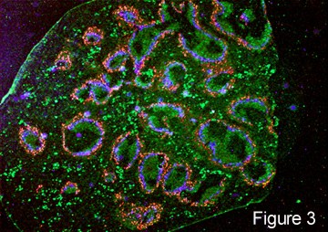
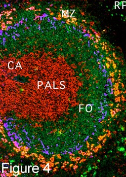
After newly formed B cells exit generative sites in fetal liver or adult bone marrow they undergo selection events that may involve interactions with self or with external antigens. Selective events can influence the phenotype and functional characteristics of B cells. B cell receptor-mediated events also influence lymphoid organs localization as marginal zone B cells in the spleen, as follicular (B-2 cells), as well as B-1 cells in the peritoneal and pleural cavities. [] fluorescence micrograph spleen, fm high power in which T cells form periarteriolar lymphocyte sheath (PALS) (red) and B-2 cell follicles (green) []

B-1 cells are the first B cells produced in the fetus, and in adults are located primarily in the peritoneal and pleural cavities. B1 cells are believed to operate in the innate response to infection by viruses and bacteria, and usually show preferential responses to T cell-independent antigens. The diversity of B-1 lymphocytes is attributed to their recombinatorial recombination, in which there is a preferential recombination between D-proximal VH gene segments. B-1 lymphocytes express (polyspecific) IgM in greater quantities than they express IgG, and the ability of B1 cells to respond to isotype switch commitment factors such as interleukin-4 may be secondary to their production of IgM. B-1 cells express CD5, which binds to CD72 to mediate B cell-B cell interactions.

B-2 cells are conventional B lymphocytes that are produced postnatally (unlike fetal B-1 cells) and are replaced from the bone marrow.

Plasma B lymphocytes are committed to production of copious amounts of monoclonal antibodies.

Memory B lymphocytes are long-lived, stimulated B lymphocytes that are primed for rapid response to a repeated exposure of the priming antigen. Memory B cells are generated in lymphoid tissue after B cell activation/proliferation and reside in the bone marrow, lymph nodes, and spleen. High affinity surface immunoglobulins enable their activation by lower levels of cognate antigen than are naïve B cells.

NK cells are differentiated from killer T cells. NK, natural killer cells constitute a corps of circulating lymphocytes that are constitutively specialized to attack cancerous cells and virus infected cells. Preprogramming for target recognition, coupled with the absense of need for backup by a clone of identical cells, renders NK cells capable of rapid (innate) response to pathogens. NK attack involves the exocytosis of cytoplasmic granules containing perforin and granzymes. Perforin forms pores in the plasma membrane of attacked cells through which serine-protease granzymes enter, cleaving caspase precursors and triggering apoptosis.

Individuals inherit multiple, polymorphic genes for NK receptors, so the assemblage of NK receptors differs between individuals. NK cells carry two forms of surface receptors:
● killer inhibitory receptors (KIRs) transmit an inhibitory signal when they encounter class I MHC molecules on a cell surface. (By contrast, T cells only recognize antigens that are presented by a MHC molecule.)
● activating receptors, which activate the NK cell upon binding to a target cell

Viral infection often causes suppression of MHC expresion, leading to a reduction of inhibition of NKs by its killer inhibitory receptors. This double negative renders the virus infected cell a target for killing by NK cells.

"About 85% of peripheral B cells are phenotypically mature and display first-order exponential kinetics defined by a half-life of 5-6 weeks, whilst the remainder are short-lived with a life span of several days."[s]

[] tem plasma cell [] micrograph macrophage surrounded by normal plasma cells [] micrograph macrophage & plasma cells []

▼ activation : BCRs : CDRs : helper T cells : life-span B cells : lymphopoiesis : naïve B cells : surface-immunoglobulins : surface receptors : VDJ recombination ▼

Tables  Fc receptors  Immune Cytokines  Immunoglobulins  Cell Adhesion Molecules  Cell signaling  Receptor Tyrosine Kinases (RTKs)  Receptor Signal Transduction  Second Messengers 

▲ Top ▲

tags

class-switch recombination

Class-switch recombination involves isotype switching between immunoglobulin classes and isotypes.

Small resting B lymphocytes initially producing IgM antibodies. The diverse antibody repertoire achieved early in B-lymphocyte development results from VDJ recombination of gene segments to produce unique heavy- and light-chain variable (V) Ig regions. The variable regions encode the antigen binding sites of antibody receptors expressed on the surface of naïve B lymphocytes and their clonal progeny. The low affinity of naïve VDJ generated antibodies is compensated for by the high affinity of secreted IgM, in which the prototypical four-chain Ig structure is combined into pentamers or hexamers. Following encounter with antigen, B cells become activated to isotype switch from IgM to other Ig classes, including high-affinity IgG and IgA antibodies required to inactivate toxins, neutralize viruses, and promote the clearance of microorganisms.

Ligation of antigen by cognate B lymphocytes accompanied by costimulation by helper T lymphocytes, activates B lymphocytes, which enter the germinal centers of peripheral lymphoid organs to become centroblast B cells. Within the germinal center, secondary antibody diversification is brought about through somatic hypermutation (SHM) and/or gene conversion (GC) of the V region to generate high-affinity antigen binding sites. (SHM is the predominant mechanism in mice and humans, whereas GC occurs in chickens and some other species.)

Within a particular centroblast B cell in the germinal center, the heavy-chain variable (V) regions encoding the antigen binding sites are rearranged down the chromosome through class-switch recombination (CSR). Then they can be expressed with one of the constant (C) region genes to perform many different effector functions having been released into the circulation.

This isotype class switch distributes a particular variable region to different constant immunoglobulin regions. Each constant region mediates a specialized effector function, and switching permits adaptive guidance of antibodies. Creating a new heavy chain requires loop-out and deletion of DNA between switch regions, employing transcription of the switch regions. Requisite switching factors include activation-induced cytidine deaminase and components of general DNA repair, including base excision repair (UNG2), mismatch repair, and double-strand break repair.[r1]

'Individuals, such as those with hyper-IgM syndrome (HIGM), who lack the ability to make such high-affinity IgG and IgA antibodies, are unable to combat bacterial and viral infections and usually die at a young age.[s]

Tables  Fc receptors  Immune Cytokines  Immunoglobulins

[r1] DNA acrobats of the Ig class switch. Wang CL, Wabl M. J Immunol. 2004 May 15;172(10):5815-21. [Free Full Text Article]

[s] The generation of antibody diversity through somatic hypermutation and class switch recombination. Li Z, Woo CJ, Iglesias-Ussel MD, Ronai D, Scharff MD. Genes Dev. 2004 Jan 1;18(1):1-11. [Free Full Text Article]

tags