Cancer

Cancers arise when cells escape normal controls on cellular proliferation.

Cancer is not a single disease, rather the term encompasses a group of conditions that share the characteristic process of uncontrolled proliferation of cells that are typically capable of local infiltration into other tissues (invasion). This propensity for invasion and migration is associated with the capacity of malignant tumors to metastasize to sites distant from the point of origin.

This propensity for invasion and metastasis is a critical feature that distinguishes malignant tumor from benign tumors. Benign tumors evidence as local overgrowth, but fortunately have minimal or no propensity for tissue infiltration and metastasis.

Cancers are named for the tissue/organ in which they originate. However, the tissue type of cancers from a particulary organ can vary, and even cancers of the same tissue type can vary considerably in degree of undifferentiation, sensitivity to chemotherapeutic agents, growth rate, invasiveness, and metastatic potential.

Alteration of a gene that usually controls cell growth can promote the uncontrolled growth characteristic of cancer. Proto-oncogenes are the normal forms of dominant genes that function in the various signal transduction cascades involved in regulation of cell growth, proliferation and differentiation – and the malignantly transformed versions of proto-oncogenes are termed oncogenes.

For example, mutation in a proto-oncogene, such as a gene that encodes an intracellular signaling protein that is normally activated only by extracellular growth factors, converts the proto-oncogene into an oncogene. The malignantly transformed oncogene encodes an altered form of the signaling protein that now behaves as though activated even in the absence of controlled growth factor binding. The malignant cell line has escaped normal gene regulation and cell cycle control mechanisms and exhibits unchecked proliferation.

There are many excellent sites with information for those affected by cancer, so the purpose of this site, in conjunction with the companion sites, is an exploration of the cell and molecular biology of malignancy.

synonyms : cancer, tumor, malignancy, neoplasm; cancerous, tumorous, malignant, neoplastic; cancer, neoplasia, oncology.

related items ¤¤ adenoviruses ¤ amplification ¤ carcinogenesis ¤ c-Fos ¤ c-Jun ¤ c-Myc ¤ c-Sis ¤ estrogen receptors ¤ gene amplification ¤ genetic predispositon ¤ HBV ¤ HIV ¤ HPV ¤ HTLV-I ¤ immune evasion ¤ irradiation ¤ malignant transformation ¤ metastasis ¤ mitogens ¤ mutagens ¤ MYC ¤ mutations ¤ neoplasia ¤ neoplastic mutations ¤ NF-κB ¤ non-mutagenic carcinogens ¤ oncogenes ¤ p53 ¤ proliferation ¤ proto-oncogenes ¤ radiation ¤ Ras ¤ Rb ¤ retroviral mechanisms of carcinogenesis ¤ retroviruses ¤ signaling molecules ¤ SRC genes ¤ T-antigens ¤ TP53 ¤ tumor antigens ¤ tumor suppressors ¤ tumorigenic viruses ¤ viral carcinogens ¤ v-Fos ¤ v-Sis ¤ v-Myc ¤
cancer staging

Diagnostic evaluation of malignancy includes determination of the cancer's pathology (tissue type, organ of origin) in addition to staging of the cancer to determine degree of local, regional, and distant spread. Clinical and pathological staging is important to decisions concerning therapy, and to estimating prognosis (a statistical measure).

The stage of a cancer is influenced by:
¤ the carcinoma's biological aggressiveness
¤ time elapsed before clinical/pathologic diagnosis – c, cs = clinical; p, ps = pathological
¤ time elapsed before/since institution of anti-cancer treatment
¤ the tumor's sensitivity to cytotoxic therapies

Cancers commence as in situ colonies of cells that have escaped normal cellular controls. Local overgrowth of the primary tumor (localized) is followed by local tissue infiltration, and ultimately by malignant penetration of adjacent tissues (regional), blood vessels or lymphatics, with shedding and transport of malignant cells and ultimate colonization of distant organs (distant, secondary tumors, metastases).

Staging systems reflect this biological progression and the tissue type. The system of staging employed will depend, to some extent, upon the specific form of cancer involved – whether it is a solid tumor or hematologic, whether it belongs to a group staged by a specific system, such as the Ann Arbor staging classification that is commonly employed to stage lymphomas.

Cell type and grade is used to stage carcinomas of the brain and spinal cord.

TNM systemTumor, Node, Metastasis
T refers to the primary (solid) tumor – X (cannot be evaluated), 'is' (in situ), 0 - 4 (size/extent)
N refers to regional lymph node involvement – X, 0-4 (extent)
M refers to metastasis – X, 0 = no metastasis, 1 = metastasis

Ann Arbor System (lymphomas)
Stage I – single region, typically a single lymph node and the surrounding area
Stage II – two regions on same side of diaphragm, an affected lymph node or organ within the lymphatic system and a second affected area
Stage III – both sides of diaphragm, including organ or area adjacent to lymph nodes or spleen
Stage IV – diffuse or disseminated involvement of one or more extralymphatic organs, including any involvement of the liver, bone marrow, or nodular involvement of the lungs.

Modifications to Ann Arbor
A = absence of constitutional symptoms
B = presence of constitutional symptoms (night sweats, fevers, unexplained weight loss of >10%)
E = "extranodal" (not in the lymph nodes) or spread from lymph nodes to adjacent tissue
X = largest deposit if >10 cm large ("bulky disease"), or mediastinum is wider than 1/3 of the chest on a chest X-ray

"Staging: Questions and Answers" at the National Cancer Institute
International Union Against Cancer, TNM

carcinogenesis

Carcinogenesis involves damage-induced genetic alterations (mutations) that produce cancers. Mutagenesis causes genetic alterations that may, or may not, result in cancer.
Tables  Malignant Transformation  Oncogenes Proto-oncogenes 

Many of the most powerful biological regulators of cell growth and proliferation are encoded by unstable mRNAs, which are targeted for rapid degradation by the cell. The loss of rapid degradation of these growth-promoting mRNAs can result in oncogenic transformation of the cell. Targeted degradation of proto-oncogene mRNAs and short-lived cytokines is controlled both by an AU-rich element (ARE) located in the 3' noncoding region, and by several proteins that bind the ARE sequence. Activation of the ARE for mRNA decay involves cotranslation of the mRNA by ribosomes, and employs the ubiquitin-proteasome pathway.[s]

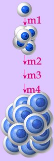
Carcinogenesis typically results from a series of mutations that affect regulation of proliferation.
m1: inactivation of a tumor suppressor gene results in cell proliferation
m2: mutation inactivates a DNA repair gene
m3: mutation of a proto-oncogene generates an oncogene
m4: mutation inactivates more cancer suppressor genes, resulting in cancerous proliferation

Carcinogenic agents include:
Mutagenic carcinogens
Non-mutagenic carcinogens
Irradiation
Viruses (tumorigenic viruses) Transforming retroviruses and DNA tumor viruses encode oncogenes.
Genetic predisposition (Table of Hereditary Cancers)

Mutagenic carcinogens: ‘genotoxic’ carcinogens are DNA reactive and induce DNA damage. Tobacco smoke is probably the most notorious mutagenic carcinogen, producing, in addition to cardiovascular damage, cancers of the head and neck, lung, and bladder.

Non-mutagenic carcinogens: ‘non-genotoxic’ carcinogens are reported to have have significantly higher computed octanol/water partition coefficients than mutagenic carcinogens, suggesting that their ability to induce tumors may be associated with membraneous receptor sites and/or a longer residence time in the animal [r]. Estrogen can promote the growth of some breast cancers.

Irradiation:

Viruses: transforming retroviruses carry oncogenes mutated from cellular genes that are involved in mitogenic signaling and growth control. DNA tumor viruses encode oncogenes of viral origin that are essential for viral replication and cell transformation; viral oncoproteins complex with cellular proteins to stimulate cell cycle progression and led to the discovery of tumor suppressors. Viral systems support the concept that cancer development occurs by the accumulation of multiple cooperating events.[s]

Genetic predisposition: a variety of inherited genetic abnormalities render affected individuals more prone to malignancy. For example, hereditary non-polyposis colon cancer (HNPCC) is a form of colon cancer frequently associated with defects in the genes encoding MSH2 (about 35% of identified gene-defect cases) and MLH1 (about 60% of identified gene-defect cases). HNPCC is characterized by early age of onset and autosomal dominant inheritance with high penetrance. (Table of Hereditary Cancers)

Cancercarcinogenesisoncogenesproliferationretrovirusessignaling moleculestumorigenic virusessite map

Tables  Apoptosis vs Necrosis  Apoptosis  Cell Adhesion  Cell signaling  Malignant Transformation  Oncogenes Proto-oncogenes  Regulatory Proteins Sequences  Table of Hereditary Cancers .

Top