metastasis

Metastasis is cell motility run amok.

Malignant tumors exhibit not only uncontrolled proliferation and local invasion, but the ability to set up distant colonies. Growth and survival of metastatic tumor cells depend upon angiogenesis and the ability of tumor cells to evade detection by the immune system.

Metastasizing cells escape normal cellular adhesion mechanisms and shed from the primary tumor. Local invasion can enable the 'escapee' cells to penetrate lymphatics and/or blood vessels. Local infiltration of lymph nodes is associated with an increased likelihood of metastasis, so determination of whether or not lymph nodes are involved is important in cancer staging.

Having been transported via circulation of lymph or blood, the malignant cells invade distant tissues where they establish focal colonies of proliferating cells (secondaries). Metastatic cancers are named for the tissue in which the primary tumor originated, for example, breast cancer metastatic to lung or bowel cancer metastatic to liver. Tumors originating in certain tissues often display a propensity for metastasis to specific tissues and organs.

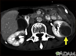
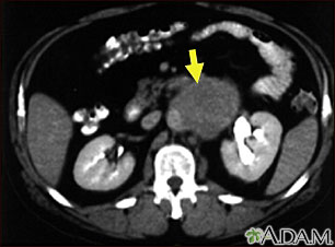
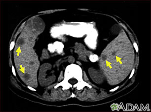
[links: images: CT scans: Kidney metastases : Liver metastases : Lymph node : Spleen metastasis ]

Top

mitogens

A Mitogen, or somatomedin, is any molecules that stimulates a cell to divide.

Most mitogens are proteins, and they stimulate signal transduction pathways that utilize mitogen activated protein kinases. Mitogens include cytokines, growth factors, hormones, neurotransmitters, cellular stress proteins, and cell adhesion ligands. For example, antigen stimulation of cell adhesion immunoglobulins triggers mitosis in B cells.

Mitogen activated protein kinases (MAP kinases) act as switch kinases that transmits information of increased intracellular tyrosine phosphorylation to that of serine/threonine phosporylation. MAPK-activated protein kinases (or MKs; formerly MAPKAP kinases) respond to mitogenic and stress stimuli through proline-directed phosphorylation and activation of the kinase domain by extracellular signal-regulated kinases 1 and 2 and p38 MAPKs.(ffta)

The signaling cascade is:
mitogen → MAPKK kinase (MAPKKK) → MAPK kinase (MAPKK)MAP kinase (MAPK) → signaling

Among the substrates of ERK are the members of the p90 ribosomal S6 kinase (RSK) family of serine/threonine kinases (10). RSK plays an active role in nuclear signaling by phosphorylating the cyclic AMP response element binding protein (CRE-binding protein, CREB) (33), c-Fos (5), and IB (27). Phosphorylation of Bad (3, 29) and C/EBPß (4) by RSK can protect cells from apoptosis. RSK has also been implicated in cell cycle regulation. RSK phosphorylates histone H3 (25), suggesting that RSK may regulate chromatin remodeling.[s-fft]

MAP kinases are also called ERKs for extracellular-signal regulated kinases, microtubule associated protein-2 kinase (MAP-2 kinase), myelin basic protein kinase (MBP kinase), ribosomal S6 protein kinase (RSK-kinase) and EGF receptor threonine kinase (ERT kinase). Maximal MAP kinase activity requires phosphorylation of both tyrosine and threonine residues. Activators of the extracellular-signal regulated kinase family (ERKs) of MAPKs include the mitogens, Ras [fft], polypeptide growth factors PDGF, CSF-1, IGF-1, EGF insulin, PMA.

Top